Post-Reproductive Ovaries Become Immune-Like and Inflammatory in Mice
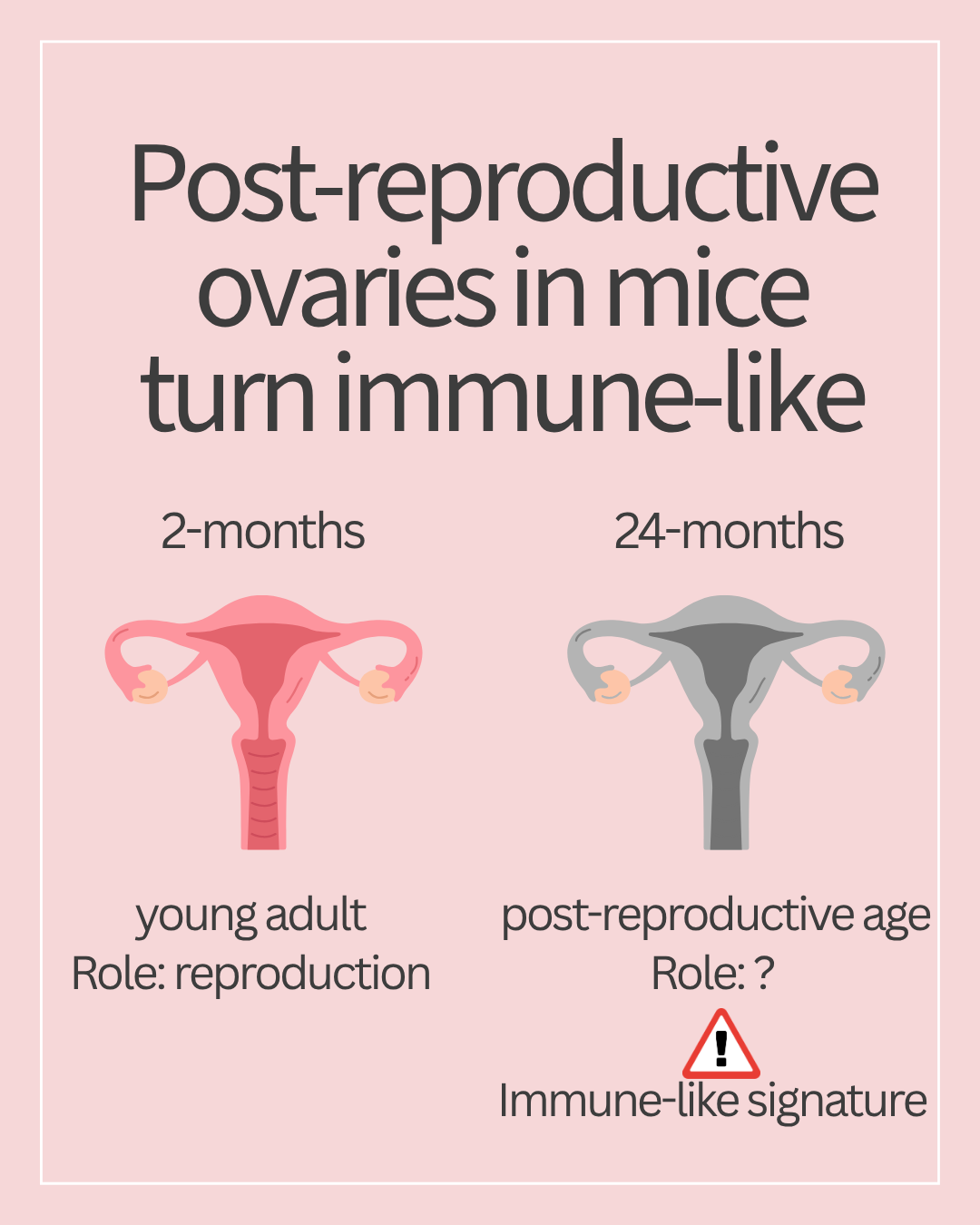
What happens to ovaries after reproduction ends? In mice, they become increasingly immune-like and inflammatory
Most research on ovarian aging focuses on what happens before reproductive function ends. These include a decline in follicle reserve, fertility, and hormone levels. Previous studies showed that ovaries turn inflammatory and fibrotic, although these studies were done in mice still within their reproductive age.
But what happens afterward? Does the ovary simply become inactive after ending its reproductive function?
A new study from the Duncan lab suggests otherwise. This team found that post-reproductive ovaries in mice become immune-like and inflammatory.
Before moving ahead, it is important to note that mice DO NOT menstruate as humans do. However, they lose ovarian follicles with age and eventually stop ovulating. Researchers call this post-reproductive state “oopause”. Studying oopause in mice can help researchers identify cellular and molecular mechanisms that can then be tested in other models and, ultimately, in human tissues.
The study
The team compared ovaries from mice at 2, 18, and 24 months of age, representing three different reproductive stages.
| Mouse age | Rough life stage |
|---|---|
| 2 months | Young adult |
| 18 months | Advanced reproductive age |
| 24 months | Post-reproductive age |
From each mouse, one ovary was examined under the microscope to look for structural changes. The other ovary was used for RNA sequencing to examine gene activity. Each group had a small number of biological replicates (n=3-4 per group).
Aging ovaries in mice have distinct structural features
Under the microscope, aging ovaries (from 18- and 24-month-old mice) showed the following changes compared to 2-month-old mice.
-
Ovarian follicles- While young ovaries contained follicles at various stages, those numbers decreased significantly by 18 months. The authors observed similar numbers between 18 and 24 months, suggesting follicle depletion was almost complete by 18 months.
-
Corpora lutea (CLs) – CLs are transient endocrine structures formed after ovulation. They were clear in 2-month-old ovaries, but not in 24-month-old ovaries, consistent with the absence of ovulation.
-
Ovarian fibrosis - Aging is associated with accumulating collagen levels leading to fibrosis. Collagen staining showed an increasing trend with age and remained high in the older cohorts.
Here, it is important to note that microscopy images provide only a snapshot in time, revealing tissue-level structural changes. But what is going on inside the cells? To examine those changes, the researchers turned to RNA sequencing.
RNA sequencing reveals post-reproductive ovary in mice acquires immune signatures
Gene activity levels largely corroborated the microscopy findings. For example, pathways involved in cell cycle and metabolism decreased with age, consistent with the loss of ovarian follicles and reproductive function.
In contrast, genes involved in immune and inflammatory activity increased. These included genes linked to innate immunity, adaptive immunity, inflammatory responses, cytokine production, and immune cell activation- all of which were higher in the older cohort. This extends observations from earlier work looking at ovaries from reproductively advanced mice.
What I found particularly interesting was what happened after most reproductive changes had already occurred (i.e., after 18 months). Especially given how similar 18- and 24-month-old ovaries were to each other compared to the 2-month-old group. Still, 230 genes differed between the two oldest groups, including changes in pathways related to immune responses and extracellular-matrix remodeling. This suggests that the ovary continues to change molecularly even after follicle depletion is largely complete.
Post-reproductive ovaries contain more immune cells
To test whether immune-related gene expression levels were reflected at the cellular and protein level, the team performed immunohistochemistry. They found age-associated increases in markers for several immune-cell populations, including T cells, macrophages, and multinucleated giant cells. They also observed increased expression of genes involved in processes such as antigen presentation and immune-cell activation. Together, these results point to an increasingly immune-like environment in the aging ovary.
Post-reproductive ovary could potentially communicate with the rest of the body
Ovaries are one of the first organs to age in the human body. Does this mean that ovarian aging impacts overall aging in the body by releasing factors that influence distant tissues? To me, this sounded like an intriguing hypothesis.
To test this hypothesis, the team looked at genes again, this time looking for those predicted to code for secreted proteins. Among them was one called Wnt7a, a gene encoding a cell signaling protein known to mediate tissue homeostasis and repair. Wnt7a gene expression levels were downregulated in ovaries from 24-month-old mice compared with the 2-month-old group. In contrast, genes encoding immunoglobulins and complement system factors, known to drive aging in other tissues, were upregulated in ovaries from 24-month-old mice compared to those from younger 2-month-old mice.
Changes in mRNA levels do not always reflect protein expression. In this study, researchers did not validate protein expression of these secreted factors. They also did not confirm whether these factors were actually secreted from the ovaries. Therefore, whether they enter circulation and influence distant organs remains unclear. For now, these results point to an interesting mechanism to explore down the road.
A few important unanswered questions
Before we look at how well these findings apply to human ovaries, a few outstanding questions remain.
-
Where did immune cells that occupy post-reproductive ovaries come from? Were they ovarian cells remodeled from the inside or from outside tissues?
-
Within post-reproductive ovaries, which immune cells contribute to the immune-like signature?
-
It is also unclear whether the predicted ovarian-secreted factors are secreted and ultimately reach distant organs to cause signs of aging.
-
A major chicken-and-egg type situation comes up: Is inflammation a cause of ovarian aging or a consequence of it?
My take
I enjoyed reading this paper; the experimental details are straightforward, and the discussion is easy to follow. They report an important finding: after reproduction in mice, ovaries develop an immune-like, inflammatory profile.
But the most provocative idea this paper puts forth is whether ovarian aging contributes to overall aging through secreted factors. That remains provocative at best, with more experiments needed to decode whether the aging ovary communicates to distant organs through some secret language (i.e., secreted factors).
Regardless, realizing that the post-reproductive ovary is a highly dynamic organ in mice can help us better understand women’s health far beyond fertility.
What is next?
Personally, I am most interested in identifying the immune cells in post-reproductive ovaries. Where did they come from? Methods like lineage tracing and single-cell sequencing can answer some of these questions.
I am equally curious to see how ovarian organoids can be used to probe the findings of this study before we ask how these findings apply to human ovaries. For example, the same lab reported developing self-organizing ovarian organoids to study aging. Could these models be used to isolate and manipulate some of the inflammatory changes observed in post-reproductive ovaries?
Reference: Converse et al., The post-reproductive ovary shifts from a reproductive to an immune-like organ, Molecular Human Reproduction, Volume 32, Issue 2, 2026, gaag038, https://doi.org/10.1093/molehr/gaag038